Images library at UTHSC helps diabetics keep sight
By Suzanne Thompson
A diagnosis of diabetic retinopathy, if made early, can be sight-saving. But as many as half of the 26 million Americans diagnosed with the condition, which involves damage to small blood vessels in the back of the eye, don’t follow through on their physicians’ recommendations to see eye-care specialists. Often it’s because the patients simply can’t afford it.
Dr. Edward Chaum, creator of a new technology that is used in conjunction with a special camera, believes if primary-care physicians serving low-income patients are able to conduct visual screenings during a routine exam, many people can be saved from a life of blindness.
“People go blind from diabetes most often because they don’t see an eye specialist in a timely fashion,” says Chaum, Plough Professor of Retinal Diseases and a professor at UT Health Science Center’s Hamilton Eye Institute (HEI). His technology allows primary-care physicians to intervene.
“What we’re trying to do is catch them early and treat them,” says Dr. James Fleming, Philip M. Lewis Endowed Professor in Ophthalmology at HEI.
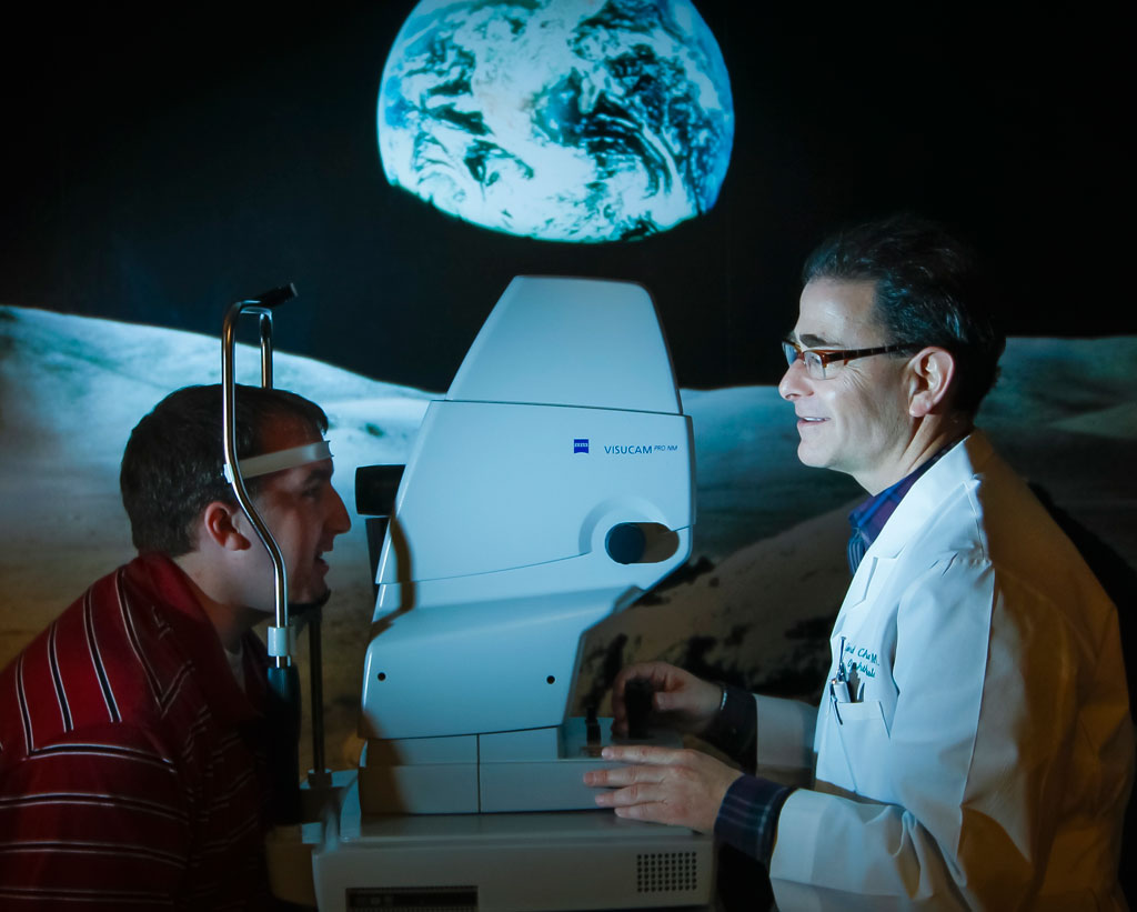
Using Content-Based Image Retrieval (CBIR), a camera photographs the retina and sends those images to a computer that compares the patient’s retinal image against those in a validated library of images. This gives physicians a quick and easy way of determining if eye disease is present and how far it has progressed. “You go into the library and determine where that image ranks compared to what images are there,” Chaum explains.
Building the library of images takes time. The more images, the better diagnostic tool CBIR becomes. “If you have large numbers of images of certain stages of the disease, then you have a very robust collection of images to compare it to, and you can be reasonably confident that it corresponds to a particular level of disease,” Chaum says.
A $125,000 seed-fund grant from Oak Ridge National Laboratory helped Chaum begin building the library. While visiting in Oak Ridge and hearing a lecture about computer chip analysis given by Kenneth Tobin, director of the Measurement Science and Systems Engineering Division, Chaum got the idea for adapting the technology to analyze retinal images. The National Eye Institute awarded grants totaling $6 million that were distributed over a six-year period. Since then, funding has been provided by numerous organizations, including the Plough Foundation and Health Outcomes Assessment Agency, operated by the federal government.
Photos taken at primary care locations are sent, via a telemedical network, to Chaum, who signs off personally on each one before adding it to the library. Currently, the library contains about 15,000 images. Using it, physicians have managed more than 6,000 patients during the last four years.
The telemedical network, called Telemedicine Retinal Imaging Analysis and Diagnosis (TRIAD), has won national awards. TRIAD has been so successful that Chaum and Tobin started a company, Hubble Telemedical, which has 13 cameras in Tennessee, Alabama and Texas. Chaum estimates the company will have cameras operating in a half dozen more states by the end of 2013.
“Basically, we’ve gone from an idea in 2004, ‘I wonder if I can use this technology that’s being applied for computer chips to change the way we manage diabetic eye diseases?’ to a commercial telemedical network and venture-backed company managing patients throughout the Mid-South in 2013,” Chaum says.